Samarium »
PDB 5lxr-7ml9 »
5zb8 »
Samarium in PDB 5zb8: Crystal Structure of the Novel Lesion-Specific Endonuclease Pfuendoq From Pyrococcus Furiosus
Protein crystallography data
The structure of Crystal Structure of the Novel Lesion-Specific Endonuclease Pfuendoq From Pyrococcus Furiosus, PDB code: 5zb8
was solved by
K.Miyazono,
T.Ito,
M.Tanokura,
with X-Ray Crystallography technique. A brief refinement statistics is given in the table below:
| Resolution Low / High (Å) | 41.12 / 2.50 |
| Space group | C 1 2 1 |
| Cell size a, b, c (Å), α, β, γ (°) | 257.354, 82.245, 116.581, 90.00, 109.13, 90.00 |
| R / Rfree (%) | 17.6 / 20.9 |
Other elements in 5zb8:
The structure of Crystal Structure of the Novel Lesion-Specific Endonuclease Pfuendoq From Pyrococcus Furiosus also contains other interesting chemical elements:
| Zinc | (Zn) | 15 atoms |
Samarium Binding Sites:
The binding sites of Samarium atom in the Crystal Structure of the Novel Lesion-Specific Endonuclease Pfuendoq From Pyrococcus Furiosus
(pdb code 5zb8). This binding sites where shown within
5.0 Angstroms radius around Samarium atom.
In total 7 binding sites of Samarium where determined in the Crystal Structure of the Novel Lesion-Specific Endonuclease Pfuendoq From Pyrococcus Furiosus, PDB code: 5zb8:
Jump to Samarium binding site number: 1; 2; 3; 4; 5; 6; 7;
In total 7 binding sites of Samarium where determined in the Crystal Structure of the Novel Lesion-Specific Endonuclease Pfuendoq From Pyrococcus Furiosus, PDB code: 5zb8:
Jump to Samarium binding site number: 1; 2; 3; 4; 5; 6; 7;
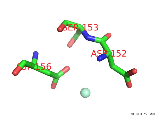
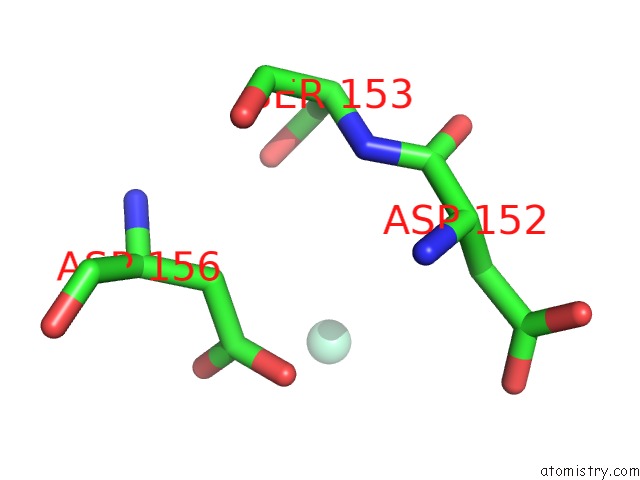
Samarium binding site 1 out of 7 in 5zb8
Go back to
Samarium binding site 1 out
of 7 in the Crystal Structure of the Novel Lesion-Specific Endonuclease Pfuendoq From Pyrococcus Furiosus

Mono view

Stereo pair view
Mono view
Stereo pair view
A full contact list of Samarium with other atoms in the Sm binding
site number 1 of Crystal Structure of the Novel Lesion-Specific Endonuclease Pfuendoq From Pyrococcus Furiosus within 5.0Å range:
|
Samarium binding site 2 out of 7 in 5zb8
Go back to
Samarium binding site 2 out
of 7 in the Crystal Structure of the Novel Lesion-Specific Endonuclease Pfuendoq From Pyrococcus Furiosus

Mono view

Stereo pair view
Mono view
Stereo pair view
A full contact list of Samarium with other atoms in the Sm binding
site number 2 of Crystal Structure of the Novel Lesion-Specific Endonuclease Pfuendoq From Pyrococcus Furiosus within 5.0Å range:
|
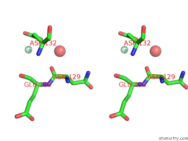
Samarium binding site 3 out of 7 in 5zb8
Go back to
Samarium binding site 3 out
of 7 in the Crystal Structure of the Novel Lesion-Specific Endonuclease Pfuendoq From Pyrococcus Furiosus
Mono view

Stereo pair view
Mono view
Stereo pair view
A full contact list of Samarium with other atoms in the Sm binding
site number 3 of Crystal Structure of the Novel Lesion-Specific Endonuclease Pfuendoq From Pyrococcus Furiosus within 5.0Å range:
|
Samarium binding site 4 out of 7 in 5zb8
Go back to
Samarium binding site 4 out
of 7 in the Crystal Structure of the Novel Lesion-Specific Endonuclease Pfuendoq From Pyrococcus Furiosus

Mono view
Stereo pair view
Mono view
Stereo pair view
A full contact list of Samarium with other atoms in the Sm binding
site number 4 of Crystal Structure of the Novel Lesion-Specific Endonuclease Pfuendoq From Pyrococcus Furiosus within 5.0Å range:
|
Samarium binding site 5 out of 7 in 5zb8
Go back to
Samarium binding site 5 out
of 7 in the Crystal Structure of the Novel Lesion-Specific Endonuclease Pfuendoq From Pyrococcus Furiosus

Mono view
Stereo pair view
Mono view
Stereo pair view
A full contact list of Samarium with other atoms in the Sm binding
site number 5 of Crystal Structure of the Novel Lesion-Specific Endonuclease Pfuendoq From Pyrococcus Furiosus within 5.0Å range:
|
Samarium binding site 6 out of 7 in 5zb8
Go back to
Samarium binding site 6 out
of 7 in the Crystal Structure of the Novel Lesion-Specific Endonuclease Pfuendoq From Pyrococcus Furiosus

Mono view

Stereo pair view
Mono view
Stereo pair view
A full contact list of Samarium with other atoms in the Sm binding
site number 6 of Crystal Structure of the Novel Lesion-Specific Endonuclease Pfuendoq From Pyrococcus Furiosus within 5.0Å range:
|
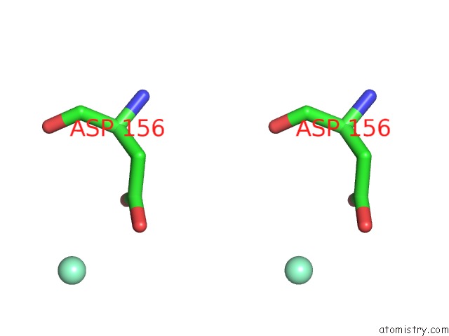
Samarium binding site 7 out of 7 in 5zb8
Go back to
Samarium binding site 7 out
of 7 in the Crystal Structure of the Novel Lesion-Specific Endonuclease Pfuendoq From Pyrococcus Furiosus
Mono view

Stereo pair view
Mono view
Stereo pair view
A full contact list of Samarium with other atoms in the Sm binding
site number 7 of Crystal Structure of the Novel Lesion-Specific Endonuclease Pfuendoq From Pyrococcus Furiosus within 5.0Å range:
|
Reference:
K.I.Miyazono,
S.Ishino,
N.Makita,
T.Ito,
Y.Ishino,
M.Tanokura.
Crystal Structure of the Novel Lesion-Specific Endonuclease Pfuendoq From Pyrococcus Furiosus. Nucleic Acids Res. V. 46 4807 2018.
ISSN: ESSN 1362-4962
PubMed: 29660024
DOI: 10.1093/NAR/GKY261
Page generated: Thu Oct 10 13:57:03 2024
ISSN: ESSN 1362-4962
PubMed: 29660024
DOI: 10.1093/NAR/GKY261
Last articles
Zn in 9MJ5Zn in 9HNW
Zn in 9G0L
Zn in 9FNE
Zn in 9DZN
Zn in 9E0I
Zn in 9D32
Zn in 9DAK
Zn in 8ZXC
Zn in 8ZUF