Samarium »
PDB 1a3c-5ktj »
5ktj »
Samarium in PDB 5ktj: Crystal Structure of Pistol, A Class of Self-Cleaving Ribozyme
Protein crystallography data
The structure of Crystal Structure of Pistol, A Class of Self-Cleaving Ribozyme, PDB code: 5ktj
was solved by
L.A.Nguyen,
J.Wang,
T.A.Steitz,
with X-Ray Crystallography technique. A brief refinement statistics is given in the table below:
| Resolution Low / High (Å) | 81.27 / 2.97 |
| Space group | I 41 2 2 |
| Cell size a, b, c (Å), α, β, γ (°) | 85.190, 85.190, 256.540, 90.00, 90.00, 90.00 |
| R / Rfree (%) | 19.6 / 25.7 |
Other elements in 5ktj:
The structure of Crystal Structure of Pistol, A Class of Self-Cleaving Ribozyme also contains other interesting chemical elements:
| Cobalt | (Co) | 10 atoms |
| Magnesium | (Mg) | 19 atoms |
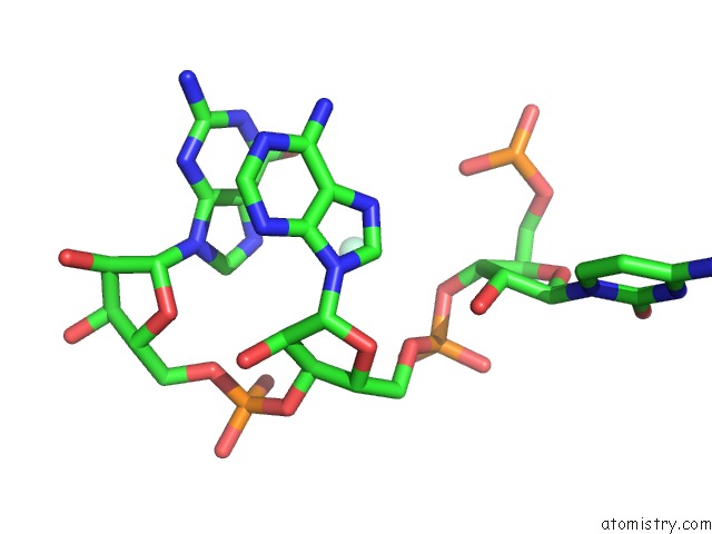
Samarium Binding Sites:
The binding sites of Samarium atom in the Crystal Structure of Pistol, A Class of Self-Cleaving Ribozyme
(pdb code 5ktj). This binding sites where shown within
5.0 Angstroms radius around Samarium atom.
In total 10 binding sites of Samarium where determined in the Crystal Structure of Pistol, A Class of Self-Cleaving Ribozyme, PDB code: 5ktj:
Jump to Samarium binding site number: 1; 2; 3; 4; 5; 6; 7; 8; 9; 10;
In total 10 binding sites of Samarium where determined in the Crystal Structure of Pistol, A Class of Self-Cleaving Ribozyme, PDB code: 5ktj:
Jump to Samarium binding site number: 1; 2; 3; 4; 5; 6; 7; 8; 9; 10;
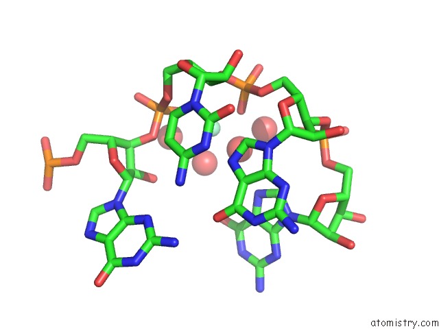
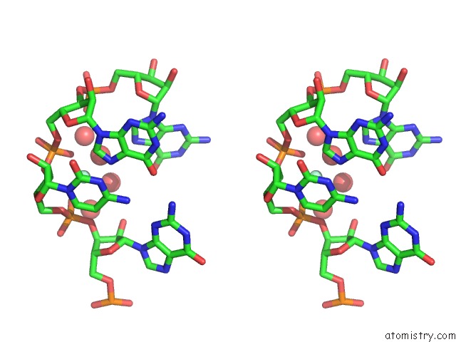
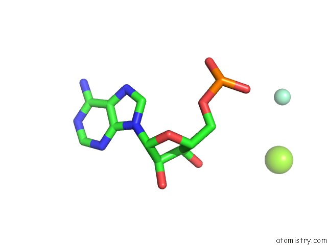
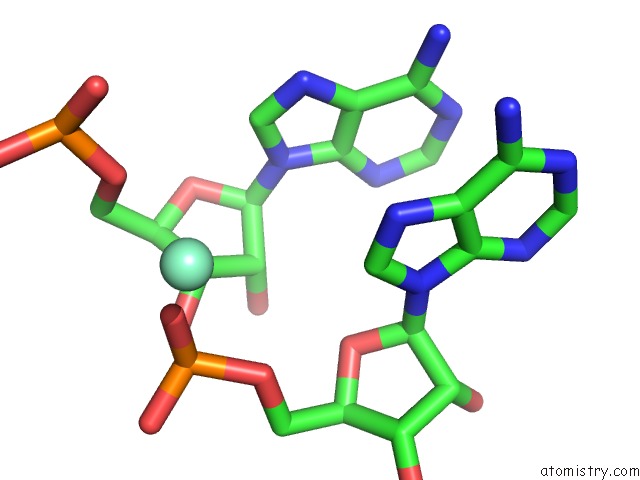
Samarium binding site 1 out of 10 in 5ktj
Go back to
Samarium binding site 1 out
of 10 in the Crystal Structure of Pistol, A Class of Self-Cleaving Ribozyme

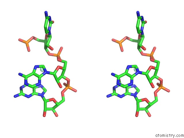
Mono view
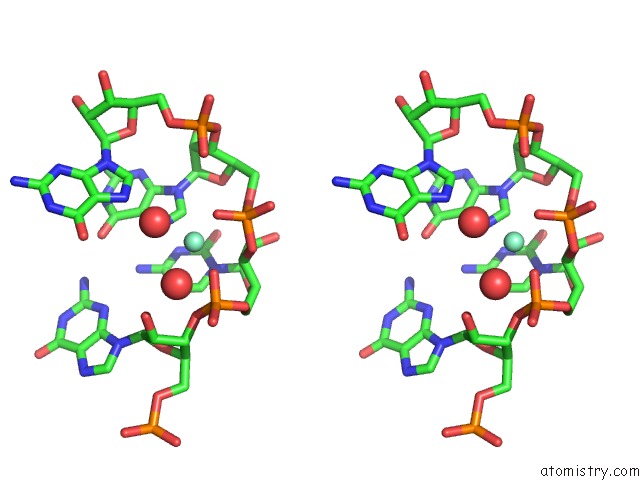
Stereo pair view
Mono view
Stereo pair view
A full contact list of Samarium with other atoms in the Sm binding
site number 1 of Crystal Structure of Pistol, A Class of Self-Cleaving Ribozyme within 5.0Å range:
|
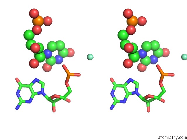
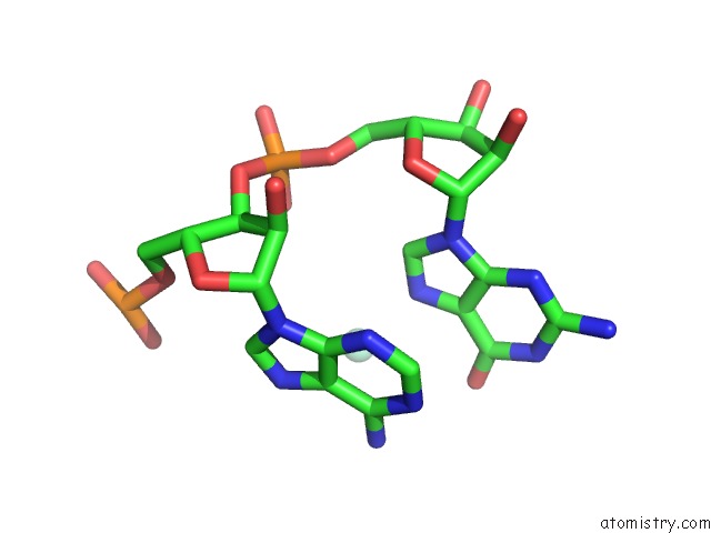
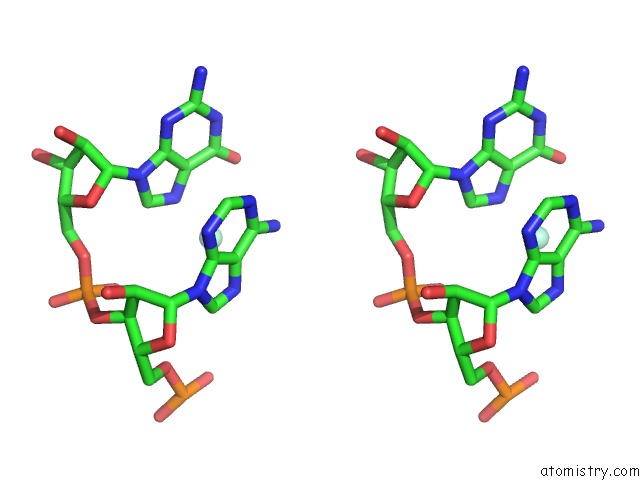
Samarium binding site 2 out of 10 in 5ktj
Go back to
Samarium binding site 2 out
of 10 in the Crystal Structure of Pistol, A Class of Self-Cleaving Ribozyme

Mono view

Stereo pair view
Mono view
Stereo pair view
A full contact list of Samarium with other atoms in the Sm binding
site number 2 of Crystal Structure of Pistol, A Class of Self-Cleaving Ribozyme within 5.0Å range:
|
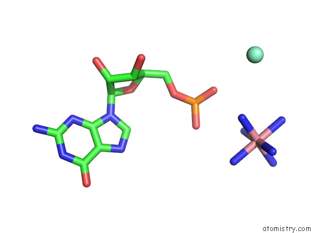
Samarium binding site 3 out of 10 in 5ktj
Go back to
Samarium binding site 3 out
of 10 in the Crystal Structure of Pistol, A Class of Self-Cleaving Ribozyme

Mono view
Stereo pair view
Mono view
Stereo pair view
A full contact list of Samarium with other atoms in the Sm binding
site number 3 of Crystal Structure of Pistol, A Class of Self-Cleaving Ribozyme within 5.0Å range:
|
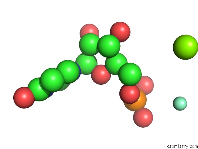
Samarium binding site 4 out of 10 in 5ktj
Go back to
Samarium binding site 4 out
of 10 in the Crystal Structure of Pistol, A Class of Self-Cleaving Ribozyme
Mono view

Stereo pair view
Mono view
Stereo pair view
A full contact list of Samarium with other atoms in the Sm binding
site number 4 of Crystal Structure of Pistol, A Class of Self-Cleaving Ribozyme within 5.0Å range:
|
Samarium binding site 5 out of 10 in 5ktj
Go back to
Samarium binding site 5 out
of 10 in the Crystal Structure of Pistol, A Class of Self-Cleaving Ribozyme
Mono view

Stereo pair view
Mono view
Stereo pair view
A full contact list of Samarium with other atoms in the Sm binding
site number 5 of Crystal Structure of Pistol, A Class of Self-Cleaving Ribozyme within 5.0Å range:
|
Samarium binding site 6 out of 10 in 5ktj
Go back to
Samarium binding site 6 out
of 10 in the Crystal Structure of Pistol, A Class of Self-Cleaving Ribozyme
Mono view
Stereo pair view
Mono view
Stereo pair view
A full contact list of Samarium with other atoms in the Sm binding
site number 6 of Crystal Structure of Pistol, A Class of Self-Cleaving Ribozyme within 5.0Å range:
|
Samarium binding site 7 out of 10 in 5ktj
Go back to
Samarium binding site 7 out
of 10 in the Crystal Structure of Pistol, A Class of Self-Cleaving Ribozyme
Mono view
Stereo pair view
Mono view
Stereo pair view
A full contact list of Samarium with other atoms in the Sm binding
site number 7 of Crystal Structure of Pistol, A Class of Self-Cleaving Ribozyme within 5.0Å range:
|
Samarium binding site 8 out of 10 in 5ktj
Go back to
Samarium binding site 8 out
of 10 in the Crystal Structure of Pistol, A Class of Self-Cleaving Ribozyme
Mono view

Stereo pair view
Mono view
Stereo pair view
A full contact list of Samarium with other atoms in the Sm binding
site number 8 of Crystal Structure of Pistol, A Class of Self-Cleaving Ribozyme within 5.0Å range:
|
Samarium binding site 9 out of 10 in 5ktj
Go back to
Samarium binding site 9 out
of 10 in the Crystal Structure of Pistol, A Class of Self-Cleaving Ribozyme
Mono view
Stereo pair view
Mono view
Stereo pair view
A full contact list of Samarium with other atoms in the Sm binding
site number 9 of Crystal Structure of Pistol, A Class of Self-Cleaving Ribozyme within 5.0Å range:
|
Samarium binding site 10 out of 10 in 5ktj
Go back to
Samarium binding site 10 out
of 10 in the Crystal Structure of Pistol, A Class of Self-Cleaving Ribozyme
Mono view

Stereo pair view
Mono view
Stereo pair view
A full contact list of Samarium with other atoms in the Sm binding
site number 10 of Crystal Structure of Pistol, A Class of Self-Cleaving Ribozyme within 5.0Å range:
|
Reference:
L.A.Nguyen,
J.Wang,
T.A.Steitz.
Crystal Structure of Pistol, A Class of Self-Cleaving Ribozyme. Proc. Natl. Acad. Sci. V. 114 1021 2017U.S.A..
ISSN: ESSN 1091-6490
PubMed: 28096403
DOI: 10.1073/PNAS.1611191114
Page generated: Thu Oct 10 13:53:34 2024
ISSN: ESSN 1091-6490
PubMed: 28096403
DOI: 10.1073/PNAS.1611191114
Last articles
Cl in 5LF4Cl in 5LF3
Cl in 5LF2
Cl in 5LF0
Cl in 5LEY
Cl in 5LF1
Cl in 5LE5
Cl in 5LDZ
Cl in 5LE1
Cl in 5LDR