Samarium »
PDB 1a3c-5ktj »
2wjd »
Samarium in PDB 2wjd: Crystal Structure of the Tyrosine Phosphatase CPS4B From Steptococcus Pneumoniae TIGR4.
Enzymatic activity of Crystal Structure of the Tyrosine Phosphatase CPS4B From Steptococcus Pneumoniae TIGR4.
All present enzymatic activity of Crystal Structure of the Tyrosine Phosphatase CPS4B From Steptococcus Pneumoniae TIGR4.:
3.1.3.48;
3.1.3.48;
Protein crystallography data
The structure of Crystal Structure of the Tyrosine Phosphatase CPS4B From Steptococcus Pneumoniae TIGR4., PDB code: 2wjd
was solved by
G.Hagelueken,
H.Huang,
J.H.Naismith,
with X-Ray Crystallography technique. A brief refinement statistics is given in the table below:
| Resolution Low / High (Å) | 29.08 / 2.80 |
| Space group | P 43 21 2 |
| Cell size a, b, c (Å), α, β, γ (°) | 88.850, 88.850, 92.310, 90.00, 90.00, 90.00 |
| R / Rfree (%) | 17.3 / 23.2 |
Other elements in 2wjd:
The structure of Crystal Structure of the Tyrosine Phosphatase CPS4B From Steptococcus Pneumoniae TIGR4. also contains other interesting chemical elements:
| Manganese | (Mn) | 3 atoms |
Samarium Binding Sites:
The binding sites of Samarium atom in the Crystal Structure of the Tyrosine Phosphatase CPS4B From Steptococcus Pneumoniae TIGR4.
(pdb code 2wjd). This binding sites where shown within
5.0 Angstroms radius around Samarium atom.
In total 2 binding sites of Samarium where determined in the Crystal Structure of the Tyrosine Phosphatase CPS4B From Steptococcus Pneumoniae TIGR4., PDB code: 2wjd:
Jump to Samarium binding site number: 1; 2;
In total 2 binding sites of Samarium where determined in the Crystal Structure of the Tyrosine Phosphatase CPS4B From Steptococcus Pneumoniae TIGR4., PDB code: 2wjd:
Jump to Samarium binding site number: 1; 2;
Samarium binding site 1 out of 2 in 2wjd
Go back to
Samarium binding site 1 out
of 2 in the Crystal Structure of the Tyrosine Phosphatase CPS4B From Steptococcus Pneumoniae TIGR4.
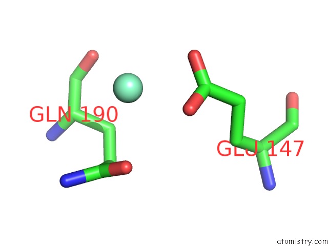
Mono view
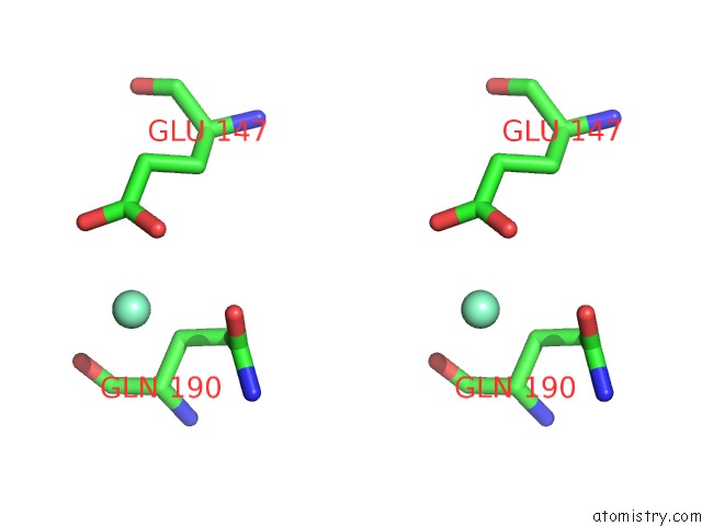
Stereo pair view
Mono view
Stereo pair view
A full contact list of Samarium with other atoms in the Sm binding
site number 1 of Crystal Structure of the Tyrosine Phosphatase CPS4B From Steptococcus Pneumoniae TIGR4. within 5.0Å range:
|
Samarium binding site 2 out of 2 in 2wjd
Go back to
Samarium binding site 2 out
of 2 in the Crystal Structure of the Tyrosine Phosphatase CPS4B From Steptococcus Pneumoniae TIGR4.
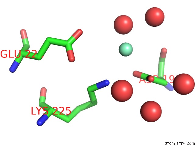
Mono view
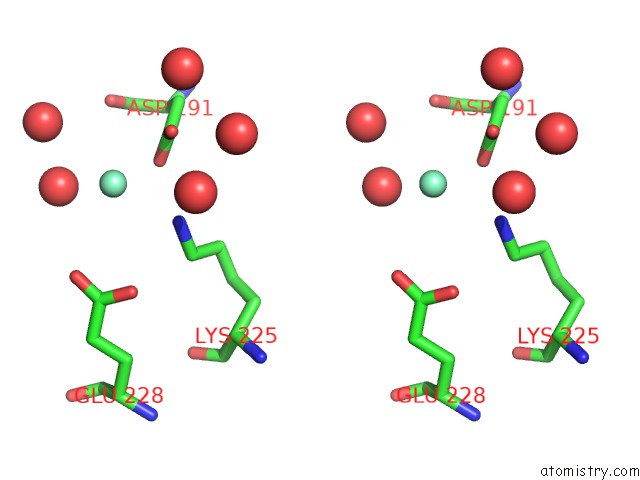
Stereo pair view
Mono view
Stereo pair view
A full contact list of Samarium with other atoms in the Sm binding
site number 2 of Crystal Structure of the Tyrosine Phosphatase CPS4B From Steptococcus Pneumoniae TIGR4. within 5.0Å range:
|
Reference:
G.Hagelueken,
H.Huang,
I.L.Mainprize,
C.Whitfield,
J.H.Naismith.
Crystal Structures of Wzb of Escherichia Coli and Cpsb of Streptococcus Pneumoniae, Representatives of Two Families of Tyrosine Phosphatases That Regulate Capsule Assembly. J.Mol.Biol. V. 392 678 2009.
ISSN: ISSN 0022-2836
PubMed: 19616007
DOI: 10.1016/J.JMB.2009.07.026
Page generated: Thu Oct 10 13:50:46 2024
ISSN: ISSN 0022-2836
PubMed: 19616007
DOI: 10.1016/J.JMB.2009.07.026
Last articles
Zn in 9MJ5Zn in 9HNW
Zn in 9G0L
Zn in 9FNE
Zn in 9DZN
Zn in 9E0I
Zn in 9D32
Zn in 9DAK
Zn in 8ZXC
Zn in 8ZUF