Samarium »
PDB 1a3c-5ktj »
2hyz »
Samarium in PDB 2hyz: Crystal Structure of An 8 Repeat Consensus Tpr Superhelix (Orthorombic Crystal Form)
Protein crystallography data
The structure of Crystal Structure of An 8 Repeat Consensus Tpr Superhelix (Orthorombic Crystal Form), PDB code: 2hyz
was solved by
T.Kajander,
A.L.Cortajarena,
L.Regan,
with X-Ray Crystallography technique. A brief refinement statistics is given in the table below:
| Resolution Low / High (Å) | 20.00 / 2.30 |
| Space group | P 21 21 21 |
| Cell size a, b, c (Å), α, β, γ (°) | 36.156, 67.695, 70.785, 90.00, 90.00, 90.00 |
| R / Rfree (%) | 21.3 / 27 |
Samarium Binding Sites:
The binding sites of Samarium atom in the Crystal Structure of An 8 Repeat Consensus Tpr Superhelix (Orthorombic Crystal Form)
(pdb code 2hyz). This binding sites where shown within
5.0 Angstroms radius around Samarium atom.
In total 3 binding sites of Samarium where determined in the Crystal Structure of An 8 Repeat Consensus Tpr Superhelix (Orthorombic Crystal Form), PDB code: 2hyz:
Jump to Samarium binding site number: 1; 2; 3;
In total 3 binding sites of Samarium where determined in the Crystal Structure of An 8 Repeat Consensus Tpr Superhelix (Orthorombic Crystal Form), PDB code: 2hyz:
Jump to Samarium binding site number: 1; 2; 3;
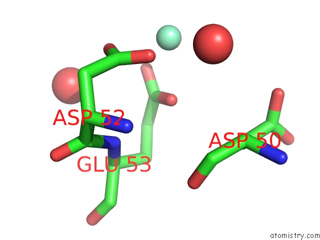
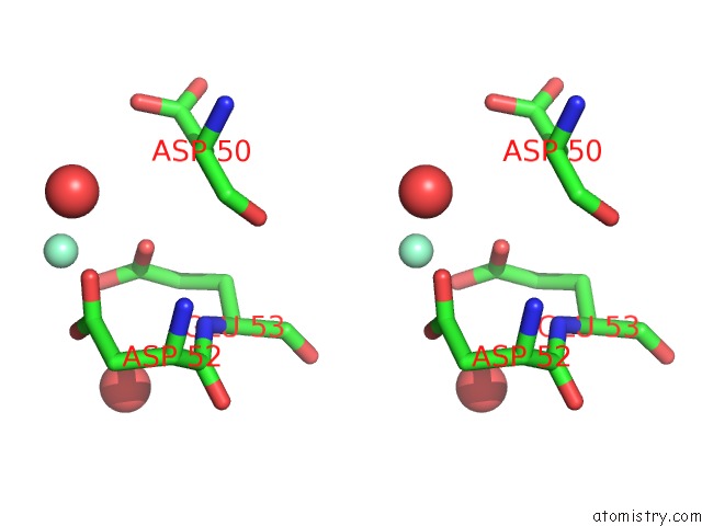
Samarium binding site 1 out of 3 in 2hyz
Go back to
Samarium binding site 1 out
of 3 in the Crystal Structure of An 8 Repeat Consensus Tpr Superhelix (Orthorombic Crystal Form)
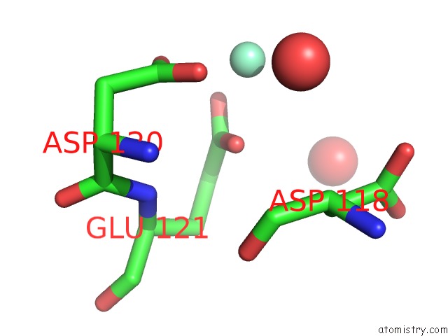
Mono view
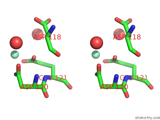
Stereo pair view
Mono view
Stereo pair view
A full contact list of Samarium with other atoms in the Sm binding
site number 1 of Crystal Structure of An 8 Repeat Consensus Tpr Superhelix (Orthorombic Crystal Form) within 5.0Å range:
|
Samarium binding site 2 out of 3 in 2hyz
Go back to
Samarium binding site 2 out
of 3 in the Crystal Structure of An 8 Repeat Consensus Tpr Superhelix (Orthorombic Crystal Form)
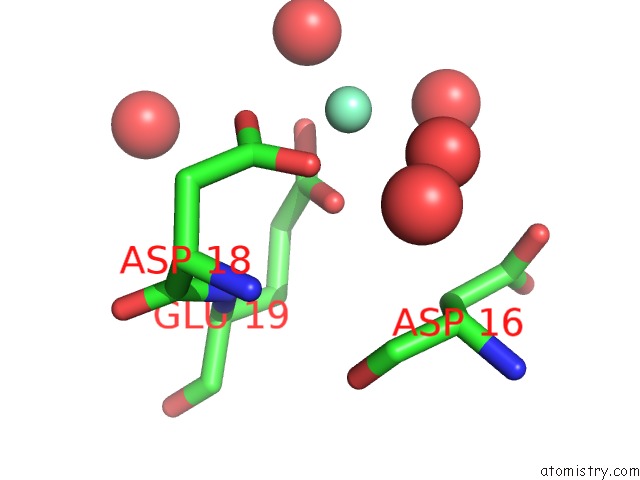
Mono view
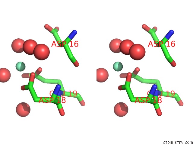
Stereo pair view
Mono view
Stereo pair view
A full contact list of Samarium with other atoms in the Sm binding
site number 2 of Crystal Structure of An 8 Repeat Consensus Tpr Superhelix (Orthorombic Crystal Form) within 5.0Å range:
|
Samarium binding site 3 out of 3 in 2hyz
Go back to
Samarium binding site 3 out
of 3 in the Crystal Structure of An 8 Repeat Consensus Tpr Superhelix (Orthorombic Crystal Form)
Mono view
Stereo pair view
Mono view
Stereo pair view
A full contact list of Samarium with other atoms in the Sm binding
site number 3 of Crystal Structure of An 8 Repeat Consensus Tpr Superhelix (Orthorombic Crystal Form) within 5.0Å range:
|
Reference:
T.Kajander,
A.L.Cortajarena,
S.Mochrie,
L.Regan.
Structure and Stability of Designed Tpr Protein Superhelices: Unusual Crystal Packing and Implications For Natural Tpr Proteins. Acta Crystallogr.,Sect.D V. 63 800 2007.
ISSN: ISSN 0907-4449
PubMed: 17582171
DOI: 10.1107/S0907444907024353
Page generated: Tue Aug 19 01:43:50 2025
ISSN: ISSN 0907-4449
PubMed: 17582171
DOI: 10.1107/S0907444907024353
Last articles
Sr in 6EELSr in 4Y1M
Sr in 6H5R
Sr in 6CAB
Sr in 6E21
Sr in 5XWG
Sr in 5V1L
Sr in 6BQ8
Sr in 4Y1J
Sr in 5WSP