Samarium »
PDB 1a3c-5ktj »
2anx »
Samarium in PDB 2anx: Crystal Structure of Bacteriophage P22 Lysozyme Mutant L87M
Enzymatic activity of Crystal Structure of Bacteriophage P22 Lysozyme Mutant L87M
All present enzymatic activity of Crystal Structure of Bacteriophage P22 Lysozyme Mutant L87M:
3.2.1.17;
3.2.1.17;
Protein crystallography data
The structure of Crystal Structure of Bacteriophage P22 Lysozyme Mutant L87M, PDB code: 2anx
was solved by
B.H.Mooers,
B.W.Matthews,
with X-Ray Crystallography technique. A brief refinement statistics is given in the table below:
| Resolution Low / High (Å) | 42.00 / 1.04 |
| Space group | C 1 2 1 |
| Cell size a, b, c (Å), α, β, γ (°) | 134.013, 50.415, 46.587, 90.00, 103.80, 90.00 |
| R / Rfree (%) | 11.7 / 15 |
Other elements in 2anx:
The structure of Crystal Structure of Bacteriophage P22 Lysozyme Mutant L87M also contains other interesting chemical elements:
| Magnesium | (Mg) | 1 atom |
| Iodine | (I) | 9 atoms |
Samarium Binding Sites:
The binding sites of Samarium atom in the Crystal Structure of Bacteriophage P22 Lysozyme Mutant L87M
(pdb code 2anx). This binding sites where shown within
5.0 Angstroms radius around Samarium atom.
In total 3 binding sites of Samarium where determined in the Crystal Structure of Bacteriophage P22 Lysozyme Mutant L87M, PDB code: 2anx:
Jump to Samarium binding site number: 1; 2; 3;
In total 3 binding sites of Samarium where determined in the Crystal Structure of Bacteriophage P22 Lysozyme Mutant L87M, PDB code: 2anx:
Jump to Samarium binding site number: 1; 2; 3;
Samarium binding site 1 out of 3 in 2anx
Go back to
Samarium binding site 1 out
of 3 in the Crystal Structure of Bacteriophage P22 Lysozyme Mutant L87M
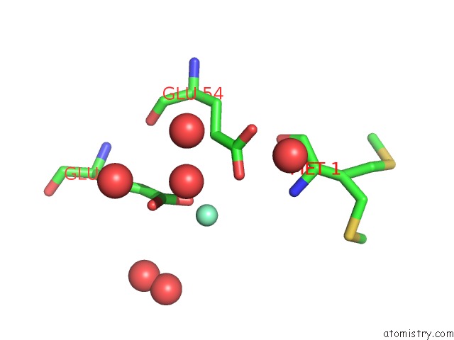
Mono view
Stereo pair view
Mono view
Stereo pair view
A full contact list of Samarium with other atoms in the Sm binding
site number 1 of Crystal Structure of Bacteriophage P22 Lysozyme Mutant L87M within 5.0Å range:
|
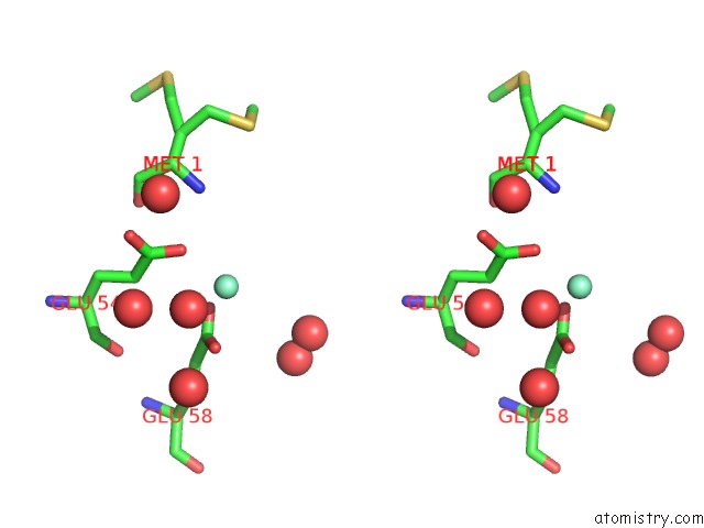
Samarium binding site 2 out of 3 in 2anx
Go back to
Samarium binding site 2 out
of 3 in the Crystal Structure of Bacteriophage P22 Lysozyme Mutant L87M
Mono view

Stereo pair view
Mono view
Stereo pair view
A full contact list of Samarium with other atoms in the Sm binding
site number 2 of Crystal Structure of Bacteriophage P22 Lysozyme Mutant L87M within 5.0Å range:
|
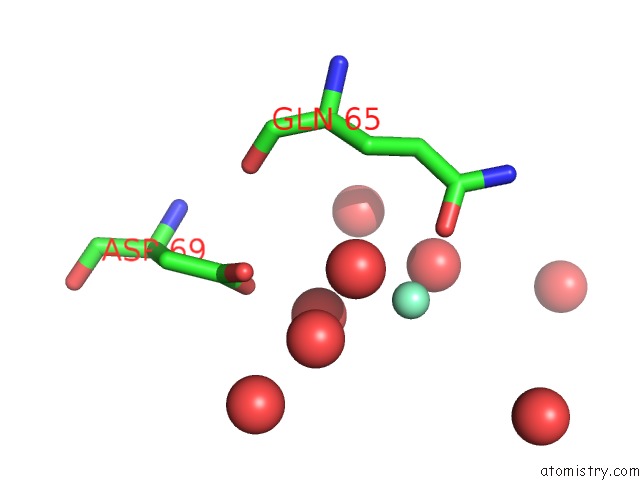
Samarium binding site 3 out of 3 in 2anx
Go back to
Samarium binding site 3 out
of 3 in the Crystal Structure of Bacteriophage P22 Lysozyme Mutant L87M

Mono view

Stereo pair view
Mono view
Stereo pair view
A full contact list of Samarium with other atoms in the Sm binding
site number 3 of Crystal Structure of Bacteriophage P22 Lysozyme Mutant L87M within 5.0Å range:
|
Reference:
B.H.Mooers,
B.W.Matthews.
Extension to 2268 Atoms of Direct Methods in the Ab Initio Determination of the Unknown Structure of Bacteriophage P22 Lysozyme. Acta Crystallogr.,Sect.D V. 62 165 2006.
ISSN: ISSN 0907-4449
PubMed: 16421448
DOI: 10.1107/S0907444905037212
Page generated: Thu Oct 10 13:49:28 2024
ISSN: ISSN 0907-4449
PubMed: 16421448
DOI: 10.1107/S0907444905037212
Last articles
Zn in 9J0NZn in 9J0O
Zn in 9J0P
Zn in 9FJX
Zn in 9EKB
Zn in 9C0F
Zn in 9CAH
Zn in 9CH0
Zn in 9CH3
Zn in 9CH1