Samarium »
PDB 1a3c-5ktj »
1soi »
Samarium in PDB 1soi: Crystal Structure of Nudix Hydrolase DR1025 in Complex with Sm+3
Protein crystallography data
The structure of Crystal Structure of Nudix Hydrolase DR1025 in Complex with Sm+3, PDB code: 1soi
was solved by
W.Ranatunga,
E.E.Hill,
J.L.Mooster,
E.L.Holbrook,
U.Schulze-Gahmen,
W.Xu,
M.J.Bessman,
S.E.Brenner,
S.R.Holbrook,
Berkeleystructural Genomics Center (Bsgc),
with X-Ray Crystallography technique. A brief refinement statistics is given in the table below:
| Resolution Low / High (Å) | 26.60 / 1.80 |
| Space group | P 41 21 2 |
| Cell size a, b, c (Å), α, β, γ (°) | 53.171, 53.171, 121.972, 90.00, 90.00, 90.00 |
| R / Rfree (%) | 21.8 / 25.1 |
Samarium Binding Sites:
The binding sites of Samarium atom in the Crystal Structure of Nudix Hydrolase DR1025 in Complex with Sm+3
(pdb code 1soi). This binding sites where shown within
5.0 Angstroms radius around Samarium atom.
In total 3 binding sites of Samarium where determined in the Crystal Structure of Nudix Hydrolase DR1025 in Complex with Sm+3, PDB code: 1soi:
Jump to Samarium binding site number: 1; 2; 3;
In total 3 binding sites of Samarium where determined in the Crystal Structure of Nudix Hydrolase DR1025 in Complex with Sm+3, PDB code: 1soi:
Jump to Samarium binding site number: 1; 2; 3;
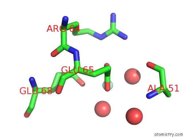
Samarium binding site 1 out of 3 in 1soi
Go back to
Samarium binding site 1 out
of 3 in the Crystal Structure of Nudix Hydrolase DR1025 in Complex with Sm+3
Mono view

Stereo pair view
Mono view
Stereo pair view
A full contact list of Samarium with other atoms in the Sm binding
site number 1 of Crystal Structure of Nudix Hydrolase DR1025 in Complex with Sm+3 within 5.0Å range:
|
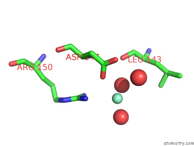
Samarium binding site 2 out of 3 in 1soi
Go back to
Samarium binding site 2 out
of 3 in the Crystal Structure of Nudix Hydrolase DR1025 in Complex with Sm+3
Mono view

Stereo pair view
Mono view
Stereo pair view
A full contact list of Samarium with other atoms in the Sm binding
site number 2 of Crystal Structure of Nudix Hydrolase DR1025 in Complex with Sm+3 within 5.0Å range:
|
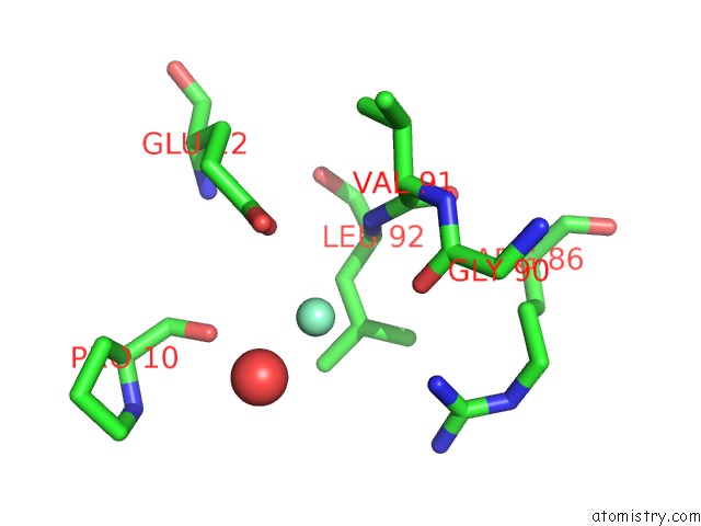
Samarium binding site 3 out of 3 in 1soi
Go back to
Samarium binding site 3 out
of 3 in the Crystal Structure of Nudix Hydrolase DR1025 in Complex with Sm+3
Mono view

Stereo pair view
Mono view
Stereo pair view
A full contact list of Samarium with other atoms in the Sm binding
site number 3 of Crystal Structure of Nudix Hydrolase DR1025 in Complex with Sm+3 within 5.0Å range:
|
Reference:
W.Ranatunga,
E.E.Hill,
J.L.Mooster,
E.L.Holbrook,
U.Schulze-Gahmen,
W.Xu,
M.J.Bessman,
S.E.Brenner,
S.R.Holbrook.
Structural Studies of the Nudix Hydrolase DR1025 From Deinococcus Radiodurans and Its Ligand Complexes. J.Mol.Biol. V. 339 103 2004.
ISSN: ISSN 0022-2836
PubMed: 15123424
DOI: 10.1016/J.JMB.2004.01.065
Page generated: Thu Oct 10 13:48:42 2024
ISSN: ISSN 0022-2836
PubMed: 15123424
DOI: 10.1016/J.JMB.2004.01.065
Last articles
Cl in 5UWXCl in 5UXB
Cl in 5UXC
Cl in 5UUZ
Cl in 5UWU
Cl in 5UVJ
Cl in 5UWR
Cl in 5UUV
Cl in 5UVF
Cl in 5UUE